
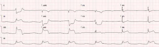
A 60 year old male with a past medical history of diabetes and tobacco use presented with epigastric pain, nausea, and vomiting. The patient was noted to have a significantly elevated blood glucose and an elevated potassium on a point-of-care chemistry at triage. The patient was immediately brought to the resuscitation team and an EKG (Figure 1) was performed:
Interpretation: Rate: 35 bpm; Rhythm: sinus bradycardia; Axis: right axis deviation (I: neg., II: pos., aVF: pos) Intervals: PR: 102, normal; QRS: 120, wide; QT: 483, mild prolongation; P-Waves: normal morphology (best seen in V5/V6); QRS Complex: right bundle branch block morphology in anterolateral leads; ST Segment/T-waves: ST elevation in II, III, aVF, & V6, with reciprocal depression in I, aVL, V1, & V2
Upon completion of this EKG, a code STEMI was immediately activated. Cardiology presented to the bedside to evaluate the patient and agreed with ED interpretation of this EKG. At this time serum laboratory testing had returned, showing an elevated glucose, potassium, and serum osmolality concerning for hyperglycemic hyperosmolar syndrome. The patient was given IV insulin and calcium prior to transfer to the cath lab.
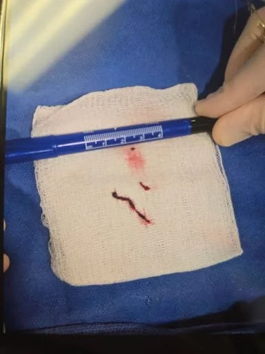
Cardiac catheterization demonstrated 100% thrombotic occlusion of the RCA. Figure 2 below shows evidence of the culprit lesion that was found in this patient.
Figure 2. Lesion isolated in cath lab with marker for scale
The patient was admitted to the cardiac ICU following the procedure and treated for both HHS and his OMI and ultimately discharged. The patient later had two subsequent admissions at which time he was treated for new onset heart failure with moderately reduced ejection fraction (EF 37%), and was doing well on most recent outpatient cardiology visit.
Discussion
The patient in this case presented with symptoms, in the setting of diabetes, we more often contribute to DKA/HHS, such as nausea, vomiting, and epigastric discomfort. Point-of-care testing further supported this presumption after the patient was brought back from triage for a significant elevation in their glucose and potassium. The EKG obtained due to the hyperkalemia demonstrated diffuse ST abnormalities and bradycardia. Careful analysis of the EKG then begs the question, without classic symptoms of OMI, are these changes secondary to hyperkalemia and metabolic abnormalities or due to acute thrombotic occlusion of a coronary artery?
First, it is important to remember that epigastric pain can be an anginal equivalent, and these patients should always receive a screening EKG. Second, it is important to always do a thorough evaluation for the cause of DKA/HHS, which includes ACS. While in this case, it is impossible to say if the RCA occlusion or the HHS came first, it is important to understand that ACS can be a trigger for DKA/HHS in at-risk populations and should be screened appropriately. Third, it is important to carefully analyze EKGs in cases of hyperkalemia for ischemia; while elevated potassium can cause both ST elevations and depressions, these are generally diffuse and do not pattern isolated coronary vascular territories or demonstrate reciprocity, as in this case.
The EKG in Figure 1 exhibits many hallmarks of an inferior OMI. Most obviously, we can see the ST segment elevation in II, II, and aVF, which are classic findings for this type of occlusion. There is also ST segment elevation in V6, which is indicative of a more extensive infarct, extending to the lateral wall. An inferior STEMI, for most patients, is caused by an RCA occlusion. However in around 18% of people, it may be secondary to a LCx occlusion. In this case, there are a few clues that point towards an RCA OMI. First, there is a proportionally larger ST elevation in lead III compared to lead II. Additionally, there is obvious ST depression in lead I. Both of these findings are characteristic of an RCA occlusion MI.
The patient was also noted to have profound bradycardia, the differential for which includes OMI, hyperkalemia, and other metabolic abnormalities. In RCA occlusion, both sinus bradycardia and bradyarrhythmias can occur due to ischemia to the SA node. Typically, the SA nodal artery is a proximal branch off of the RCA, so in extensive RCA infarcts, this branch may be affected. Despite the metabolic disequilibrium in our patient, with EKG findings suggestive of extensive RCA infarct (e.g. ST elevation in V6), high RCA OMI was likely the cause of the bradycardia in this patient. For most patients, the bradycardia resolves within the first few days and does not require pacemaker placement or further medical management.
The patient received timely transfer to the cath lab for stenting and reperfusion and had an uncomplicated recovery in the CCU. However days later, the patient experienced new onset heart failure following discharge from the hospital. This is a common complication of RCA occlusion, particularly when extensive, as in this patient.
Take-away Points:
Always obtain screening EKGs in patients with potential anginal equivalents. This includes patients >50 years old with epigastric pain.
Be sure to always dig deeper to find the root cause of presentations of DKA or HHS.
In cases of hyperkalemia, carefully investigate the electrocardiographic abnormalities to check for vascular distribution patterns and reciprocity which suggest OMI >> metabolic changes.
In patients with inferior MIs, be cognizant of potential complications, including AV blocks, bradycardia, bradyarrhythmias, and heart failure.
For most inferior MIs, the culprit lesion is going to be the RCA, however be on the look-out for clues to suggest otherwise!
References:
Burns, E. Buttner, R. Inferior STEMI. Life in the Fast Lane. Oct 8, 2024. https://litfl.com/inferior-stemi-ecg-library/
Nikus K, Birnbaum Y, Fiol-Sala M, Rankinen J, de Luna AB. Conduction Disorders in the Setting of Acute STEMI. Curr Cardiol Rev. 2021;17(1):41-49. doi: 10.2174/1573403X16666200702121937. PMID: 32614749; PMCID: PMC8142368.
Tivakaran VS, Rout P. Inferior Myocardial Infarction. [Updated 2025 Nov 7]. In: StatPearls [Internet]. Treasure Island (FL): StatPearls Publishing; 2026 Jan-. Available from: https://www.ncbi.nlm.nih.gov/books/NBK470572/
Nikus K, Birnbaum Y, Fiol-Sala M, Rankinen J, de Luna AB. Conduction Disorders in the Setting of Acute STEMI. Curr Cardiol Rev. 2021;17(1):41-49. doi: 10.2174/1573403X16666200702121937. PMID: 32614749; PMCID: PMC8142368.
Authored by Erica Dolph MD and Ari Edelheit MD