
CARDIAC TAMPONADE - CONSIDER THE PROBE
Case: A 52-year-old man with no reported PMH presents with 2 days of rapidly worsening shortness of breath that is worse when lying flat and with exertion. He endorses some vague chest pain that radiates to the neck and epigastric region.
His vitals: HR 120, BP 70/40, RR: 40, afebrile, SpO2 98% on RA
On exam he has clear lungs with obvious JVD, but no overt leg swelling or other signs of volume overload.
While the nurses get access, you consider the probe. You start with your trusty parasternal long (PSL) view and within seconds you’ve found the culprit.
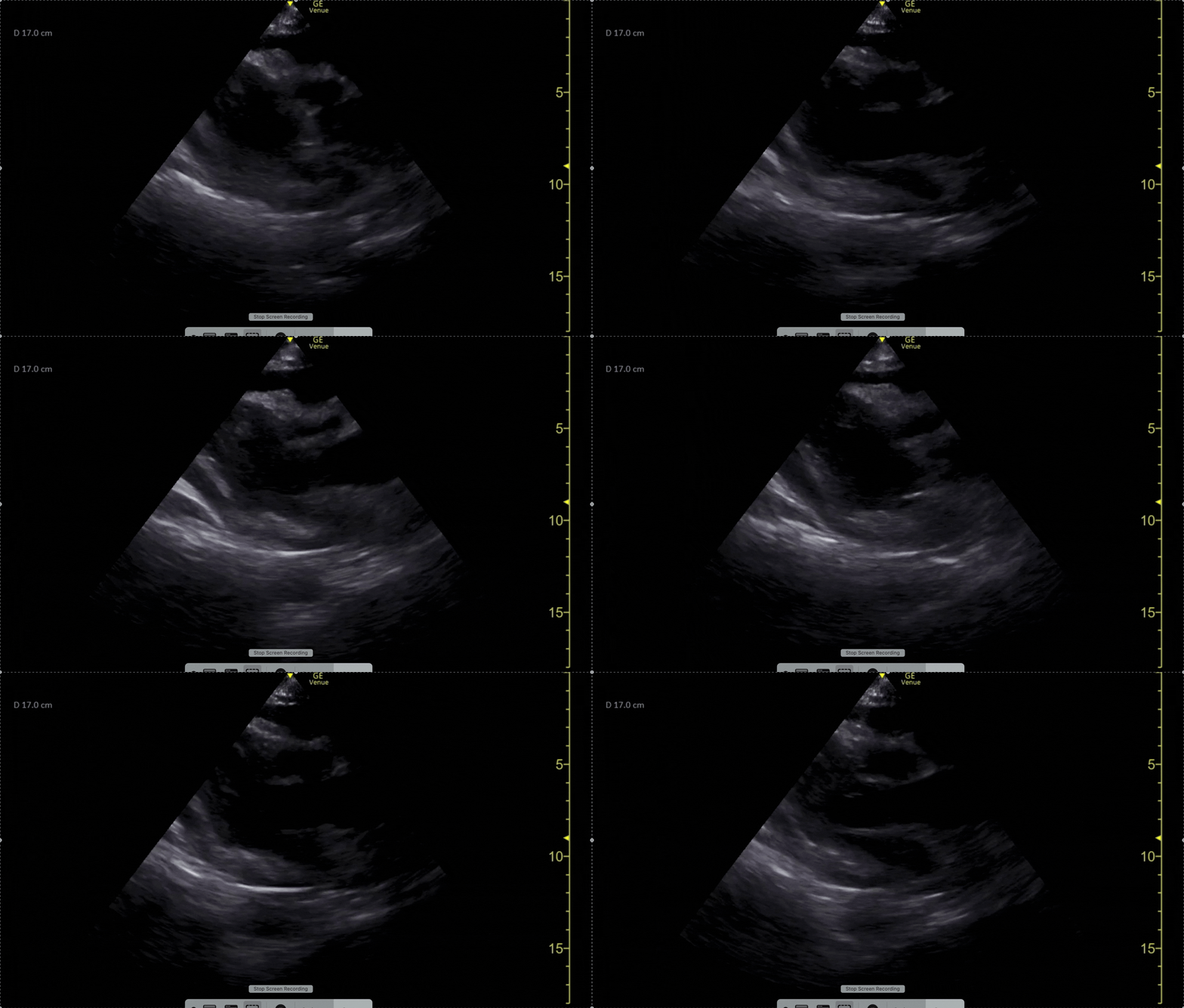
Figure 1. PSL video with circumferential effusion.
Here you see a circumferential effusion, which you confirm is in the pericardium and not a pleural effusion, as it tracks anterior to the descending aorta. So you see an effusion, but how do you know if it’s tamponade?
The size of the effusion won’t give you this answer, as a rapidly accumulating small effusion can cause tamponade, while a large but slowly progressive one may not.1
IVC
Start with your sensitive tests. Check the IVC. A plethoric IVC is the most sensitive ultrasonographic sign of tamponade (95-97% sensitivity), but it’s not very specific as this can be seen in other pathologies that elevate right sided pressures.2 The primary exception to its high sensitivity would be the hypovolemic patient, such as in trauma, where one can have tamponade with a collapsible IVC.
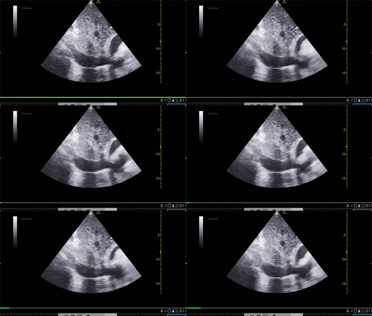
Figure 2. Video of a plethoric IVC in a patient with cardiac tamponade.
RIGHT ATRIUM
Right atrial systolic collapse is often noted before RV diastolic collapse since it’s the lowest pressure chamber. It’s highly sensitive, approaching 100% in late tamponade, but like the IVC, it’s a less specific finding.2
RIGHT VENTRICLE
RV diastolic collapse is a specific finding (75-90%), but it’s less sensitive (48-60%), so its absence doesn’t rule out tamponade. In early tamponade, one may only see collapse during expiration, when RV preload drops.2
The trick is getting the proper timing. Because systole and diastole blur together in the tachycardic patient, M-mode is your friend here. Start in the PSL view, drop the line through the RV wall, septum, and the anterior leaflet of the mitral valve, just like EPSS. You’ll notice the mitral valve trace a tall E wave (early diastole), and a smaller A wave (the atrial kick).
Identify an E wave and trace straight up. If the RV wall is dipping toward the septum during that same moment, you’ve captured diastolic collapse.
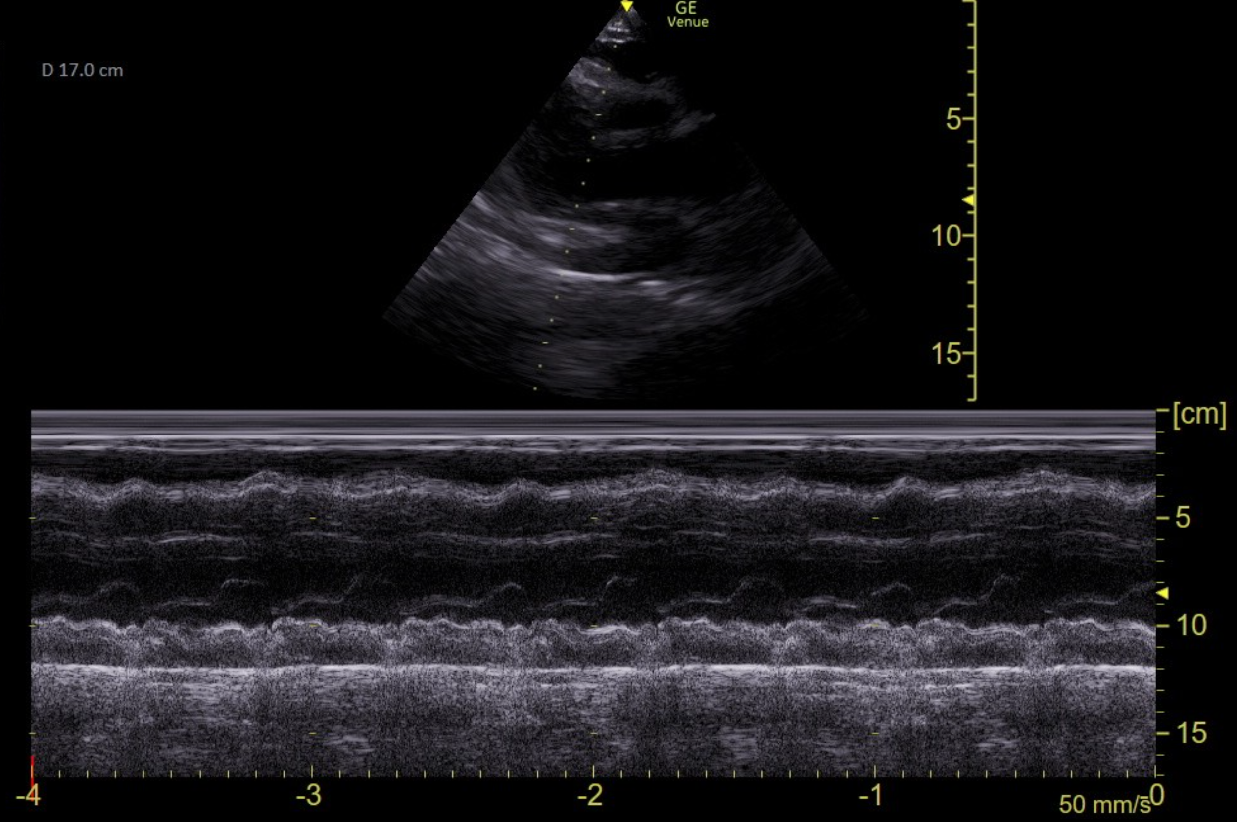
Figure 3. Here is a parasternal view of the heart in M mode, with the line tracing through the RV, septum, and mitral valve.
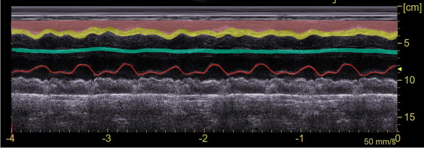
Figure 4. This is the same tracing as Figure 3. Here from bottom to top you see the mitral valve highlighted in red, the septum in teal, the RV wall in yellow, and the effusion in pink. Note the RV collapses downward toward the septum during each E wave of the mitral valve.
SONOGRAPHIC PULSUS PARADOXUS
Another specific finding in cardiac tamponade is respiratory variation in the mitral valve peak E wave inflow velocity, known as sonographic pulsus paradoxus, measured with pulse wave doppler in the A4 view. Normally the variation should be around 5% in a healthy patient, but a finding of > 25-30% inspiratory decrease is specific for tamponade.1,3
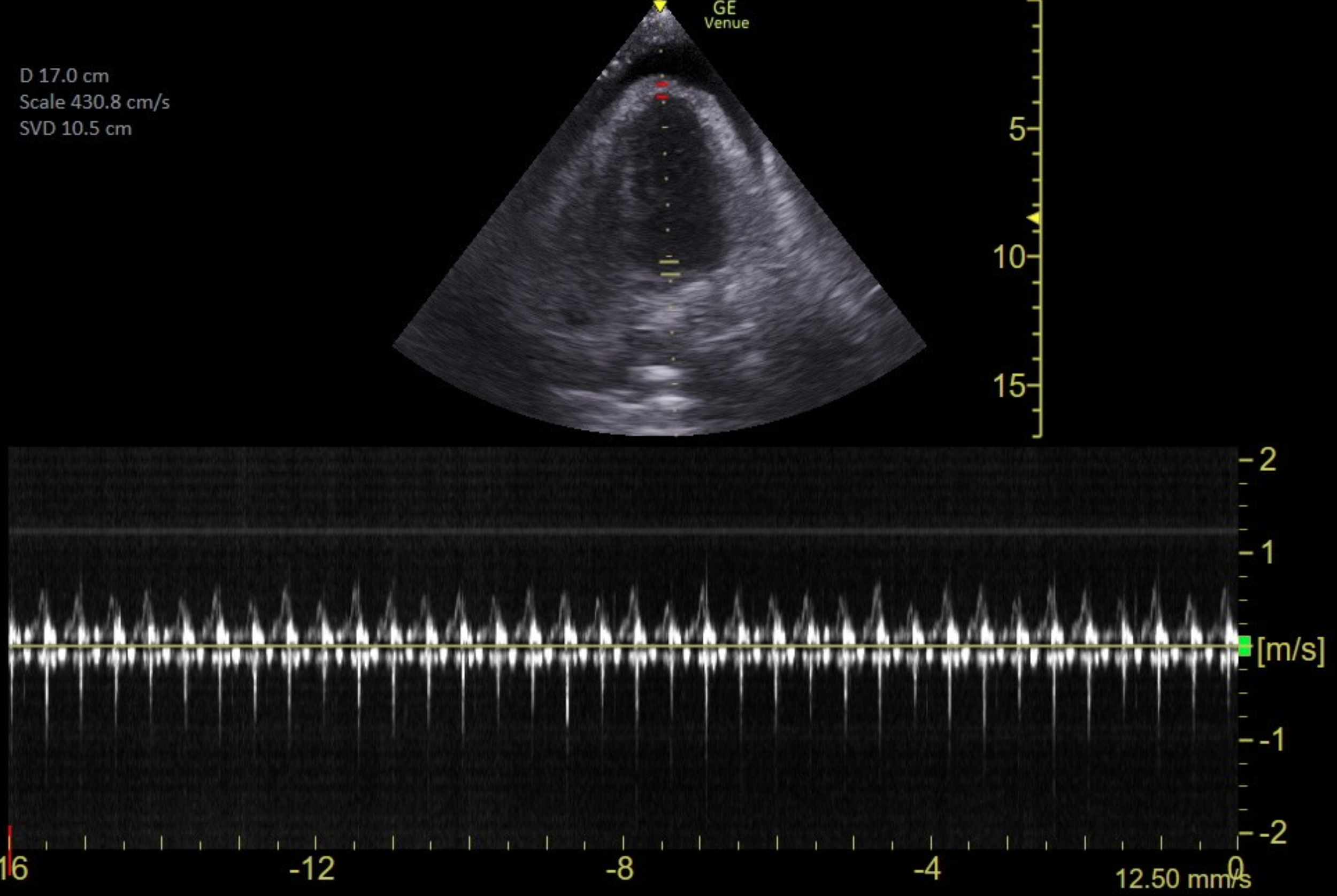
Figure 5. Pulse wave doppler at the mitral valve in the A4 view. Here there is only an approximate 20% variation in inspiratory/expiratory peak velocities, a reminder that a negative finding doesn’t rule out your diagnosis of tamponade.
CAUTION
Loculated effusions and post-surgical adhesions can muddy all of these findings, so in the wise words of the radiologist, always correlate clinically.
AIRWAY
For these patients, an intubation can kill, so avoid as much as possible. This positive pressure decreases preload while simultaneously contributing to extracardiac compression, a dangerous combination that may lead to arrest upon intubation. If it cannot be avoided, opt for hemodynamically supportive agents, such as ketamine, to avoid cutting the sympathetic drive. Once intubated, use a high respiratory rate, low tidal volumes, and minimal PEEP.2
DRAINAGE
The days of the blind pericardiocentesis are fading, and literature supports drastically lower complication rates when using ultrasound (15-20% to 0.5-3.7%, respectively).2The ideal approach is in-plane with the linear probe in the view where the effusion is the largest (typically the A4 or PSL view). Use the curvilinear probe if this provides the best view. Decrease the depth until you see only the effusion and the heart wall, and watch your needle the whole way. In the PSL approach, be careful to avoid the internal thoracic artery, running 1-3cm lateral to the sternal border.2
Using a pericardiocentesis kit is ideal (Seldinger technique). If this kit is not available, consider using a long 18g or larger angiocatheter or a central line kit, in order to leave a catheter behind.
CONCLUSION
In this case, drainage of the effusion revealed a purulent pericarditis from a liver abscess that had eroded through the diaphragm and pericardium. Cultures grew Salmonella and the patient had a great recovery after treatment.
Remember, use your descending thoracic aorta to identify pericardial effusions, the IVC is sensitive and RV diastolic collapse is specific for tamponade, practice in-plane technique and avoid intubation in these cases.
SOURCES
Boivin, Z., & Stenberg, R. T. (2024, June 4). Cardiac tamponade. American College of Emergency Physicians. https://www.acep.org/emultrasound/newsroom/may-2024/cardiac-tamponade
Alerhand, S., Adrian, R. J., Long, B., & Avila, J. (2022). Pericardial tamponade: A comprehensive emergency medicine and echocardiography review. The American Journal of Emergency Medicine, 58, 159–174. https://doi.org/10.1016/j.ajem.2022.05.001
Klein, A. L., Abbara, S., Agler, D. A., Appleton, C. P., Asher, C. R., Hoit, B., Hung, J., Garcia, M. J., Kronzon, I., Oh, J. K., Rodriguez, E. R., Schaff, H. V., Schoenhagen, P., Tan, C. D., & White, R. D. (2013). American Society of Echocardiography clinical recommendations for multimodality cardiovascular imaging of patients with pericardial disease. Journal of the American Society of Echocardiography, 26(9), 965–1012. https://doi.org/10.1016/j.echo.2013.06.023