Thoracic
Learning Objectives:
Describe the indications and limitations of POC thoracic US
Perform POCUS protocols for the detection of:
Pneumothorax
Pleural Effusion
Acute Interstitial Syndrome (CHF, ARDS, focal or multifocal pneumonia, pulmonary contusion)
Pneumonia
Identify relevant US anatomy of thoracic structures
Recognize the relevant findings and pitfalls when evaluating for thoracic pathology
Recognize the sonographic findings of tracheal and esophageal anatomy, especially in regard to EM procedures
Integrate thoracic US findings into individual patient and departmental management
Indications:
Acute pneumothorax
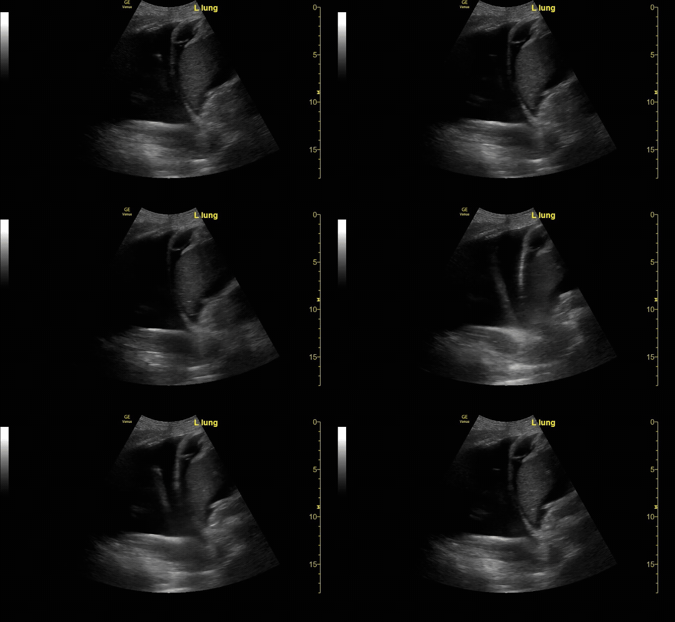
Abnormal pleural fluid collections
Presence of interstitial lung fluid (CHF, ARDS)
Extended Indications:
Identification of pneumonia, rib fractures, pulmonary fibrosis
Use POC thoracic ultrasound to guide thoracentesis or tube thoracostomy
Required Views:
Evaluate the pleural line in both lungs showing lung slide (seen here).
c/o Victoria Gonzalez, MD
M-Mode on the pleural line showing “sand & shore” indicating lung slide.
c/o Victoria Gonzalez
Thoracic ultrasound showing A-lines. Evaluate in 4 lung fields. Ensure a depth of 12-18cm.
c/o Victoria Gonzalez, MD
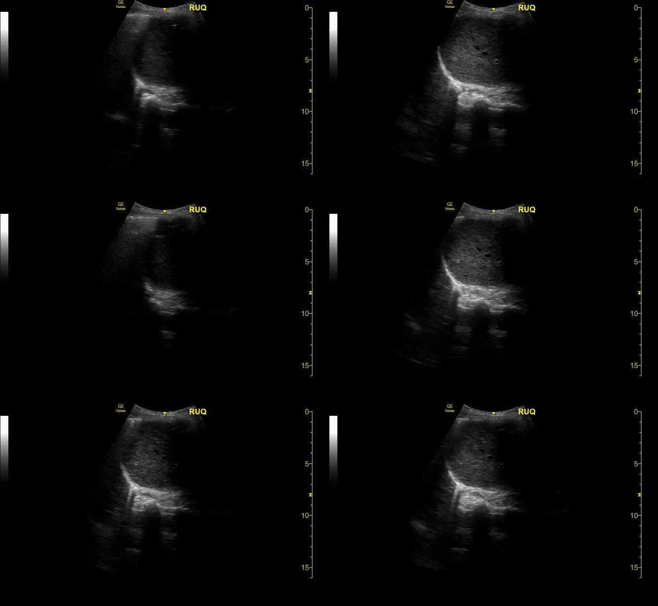
RUQ View with window curtain sign.
c/o Vishal Mittal, MD
How to Scan:
ACEP Sonoguide: Lung
POCUS101 Lung Ultrasound Made Easy
Tips/Tricks/Pitfalls:
Presence of lung slide reliably rules out pneumothorax in the area being scanned (highly sensitive)
The absence of lung slide does not always diagnose pneumothorax. Prior pleurodesis, scarring, mainstem intubation of contralateral lung, blebs, severe reactive airway disease, and bronchial obstruction can also cause this finding
A “transition point” or “lung point” where absent lung slide meets dynamic pleural sliding is highly specific for the diagnosis of pneumothorax
Remember to use enough depth (≥12 cm) when assessing the lung parenchyma
Lung ultrasound can help to differentiate a simple pleural effusion from a complex and/or loculated pleural effusion and help guide a safer thoracentesis or pigtail tube thoracostomy
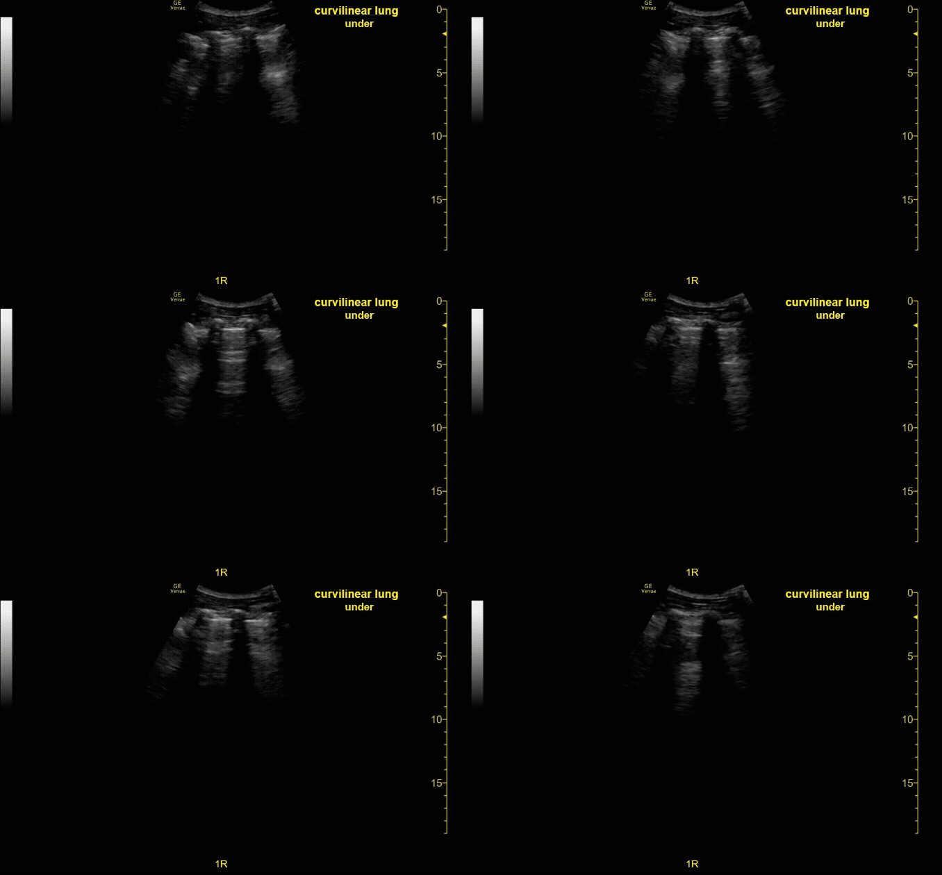
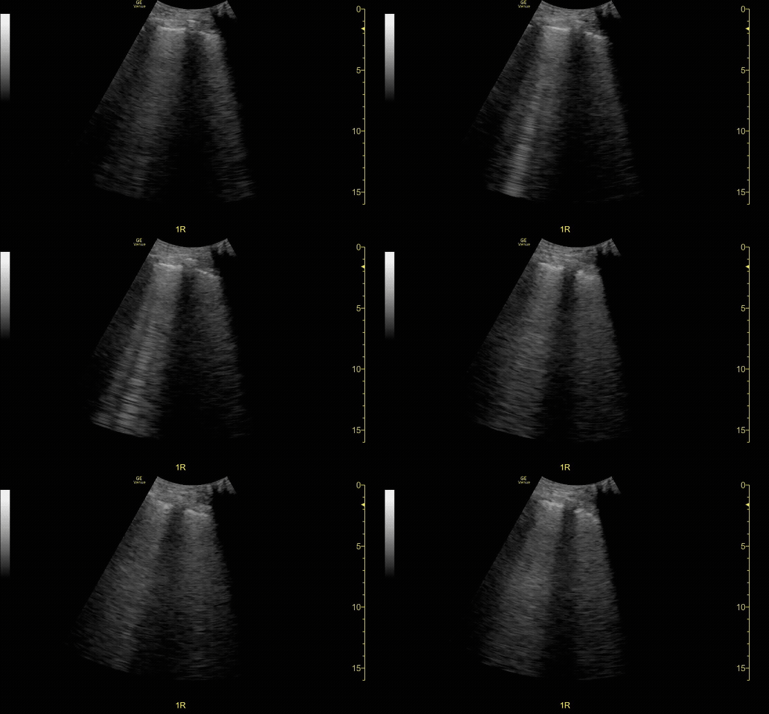
Presence of B-lines may indicate EITHER pulmonary edema or an inflammatory process while the presence of subpleural consolidation or “shred sign” indicate a focal inflammatory process
Pathology:
Pneumothorax confirmed by lung point, lung slide present to the right of the screen, no lung slide to the left.
c/o Victoria Gonzalez, MD
RUQ view showing large peural Effusion as well as ascites.
c/o Derek Hilgers, MD
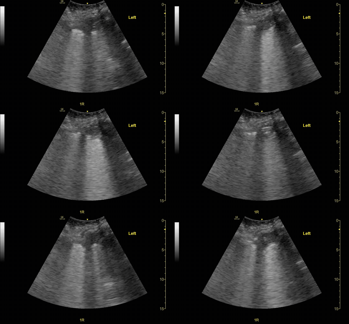
B-Lines indicating interstitial abnormalities. Ensure a depth of 12-18cm
c/o Kevin Boubouleix, MD
Thoracic ultrasound showing a shred sign (irregularity of the pleural line) indicating consolidation.
c/o Alex Alanis, MD
Key Literature:
Additional Resources:
Malin and Dawson iBook Volume 1, Chapter 5: Lung
ACEP Emergency Ultrasound Imaging Criteria Compendium Pages 16-20
Author: Susan Mari, MD (Class of 2026)
Reviewed by: David Murray, MD FPD-AEMUS


