
EFAST
Learning Objectives:
Describe the indications, clinical algorithm, and limitations of US in blunt and penetrating thoracoabdominal trauma
Perform the EUS protocol for Trauma in both primary and secondary surveys
Identify relevant US anatomy including the pleura, diaphragm, inferior vena cava, pericardium, liver, spleen, kidneys, bladder, prostate and uterus
Recognize pathologic findings and pitfalls in the evaluation of pneumothorax, hemothorax, pulmonary contusion, hemopericardium, cardiac activity, volume status, and hemoperitoneum.
Integrate Trauma EUS findings into individual patient, departmental, and disaster management
Indications:
Rapid assessment of the thoracoabdominal region for evidence of traumatic free fluid in the peritoneal, pericardial, and pleural cavities
Extended Indications:
Evaluation of solid organ injury and triage of multiple or mass casualties
Serial bedside reassessments of acute changes in patient clinical status
Required Views:
RUQ view showing liver, diaphragm, spine, and no intrathoracic fluid.
c/o Chanell Davis, MD
Pelvis in transverse view.
c//o Erica Dolph MD

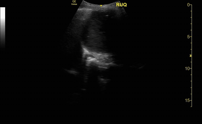
RUQ view showing diaphragm, liver (including tip), kidney, and Morrison’s pouch.
c/o Taylor Wahrenbrock, MD

Bladder in sagittal view.
c/o Erica Dolph, MD

LUQ view with diaphragm, spleen, kidney, splenorenal space and paracolic gutter.
c/o Kristina Pfieffer MD

Evaluate the pleural line in both lungs showing lung slide (seen here).
c/o Victoria Gonzalez, MD

Subxiphoid view (seen here) or parasternal long views to evaluate for pericardial fluid.
c/o Vishal Mittal, MD

M-Mode on the pleural line showing “sand & shore” indicating lung slide.
c/o Victoria Gonzalez
How to Scan:
Tips/Tricks/Pitfalls:
RUQ and LUQ structures move as the diaphragm contracts, so consider asking your patient to hold their breath
Consider slightly rotating probe counterclockwise to fit the probe better between rib spaces
In pelvis, if you are not able to get proper images, your probe is likely too superior – bladder is directly posterior to pubic bone
In LUQ, the perisplenic space is the most common spot for fluid, not the splenorenal space due to the splenorenal ligament that connects these and prevents fluid accumulation
Adequate pain control, if patient clinical status allows, can allow for improved views
If challenging/limited views despite good positioning and windows, consider pneumoperitoneum. May see the presence of abdominal A-lines
FAST does not reliably identify solid organ injuries or retroperitoneal hemorrhage
Small hemothoraces may be missed in supine position
Things that can be mistaken for free fluid: fluid in the stomach or bowel, perinephric fat (double line sign), epicardial fat pads, pericardial cysts, descending aorta
Solid organ injuries, mesenteric vascular injuries, hollow viscus injuries, and diaphragmatic injuries may likely not give rise to free fluid so could be missed by FAST
Pathology:

RUQ view showing a positive spine sign indicating free fluid in the intrathoracic cavity. Fluid also seen in the intraperitoneal space.
c/o Adam Roussas, MD

LUQ shows free fluid in the left upper quadrant around the spleen.
c/o Erica Dolph, MD

Pericardial effusion seen here with the phased array probe in subxiphoid view.
c/o Susan Mari, MD

Pneumothorax confirmed by lung point, lung slide present to the right of the screen, no lung slide to the left using linear probe.
c/o Victoria Gonzalez, MD
Key Literature:
Ma et al, How fast is the focused assessment with sonography for trauma examination learning curve?
Wilkerson, Sensitivity of Bedside Ultrasound and AP CXR for ID of PTX after trauma
Richards et al, Focused Assessment with Sonography in Trauma - What Radiologists Can Learn
Additional Resources:
Malin and Dawson iBook Volume 1 and Volume 2
Chapter 1: EFAST by Phil Craven and Mike Mallin
Chapter 22: SUSS IT by Casey Parker and James Rippey
ACEP Imaging Compendium - pg 41-46
The POCUS Atlas - 1 minute Image Review - Normal and Pathology
SAEM article with normal and pathology examples
Core Ultrasound clip bank of FAST pathology
Author: Ryan Abbott, DO (Class of 2027)
Reviewed by: Kyle Ackerman, MD