
Lower extremity DVT
Learning Objectives:
Describe the indications and limitations of EUS for the detection of deep venous thrombosis
Understand the differences between focused lower extremity venous EUS and radiology or vascular lab-performed “Duplex evaluation”
Perform EUS protocols for the detection of deep venous thrombosis of the lower extremity, including:
Vessel identification, compression, and doppler imaging of respiratory variation and augmentation
Identify relevant US anatomy of the lower extremities, including the deep venous and arterial systems, major nerves, and lymph nodes
Recognize the relevant findings and pitfalls when evaluating for deep venous thrombosis
Integrate EUS for deep venous thrombosis into individual patient management and departmental workflow
Indications:
Evaluation for acute proximal DVT in the lower extremities.
Extended Indications:
Chronic DVT
Distal DVT
Superficial venous thrombosis
Diagnosis of other causes of lower extremity pain and swelling under consideration in the evaluation of DVT such as cellulitis, subcutaneous abscess, muscle hematoma, pyomyositis, lymphadenitis, aneurysm, fasciitis, and Baker’s cyst
Required Views:
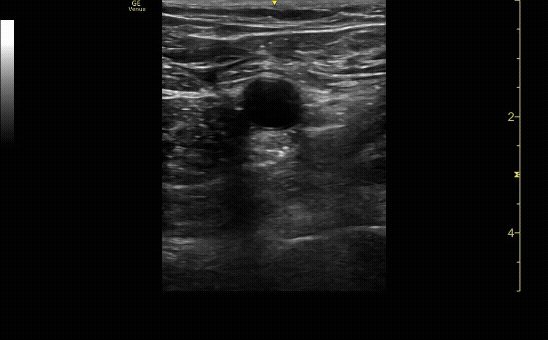
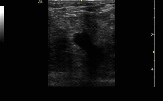
Ultrasound showing compression at the junction of greater sapphenous vein and femoral vein. Femoral artery with pulsatile flow. Be sure to show the vein before and after compression.
c/o Kyle Ackerman, MD
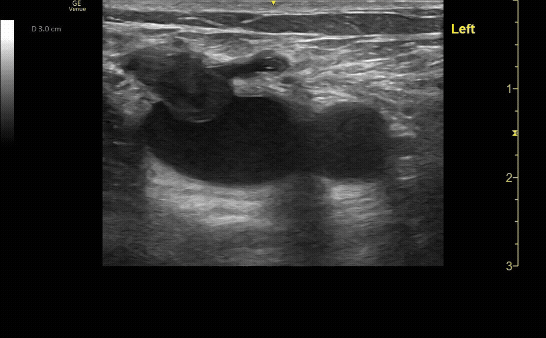
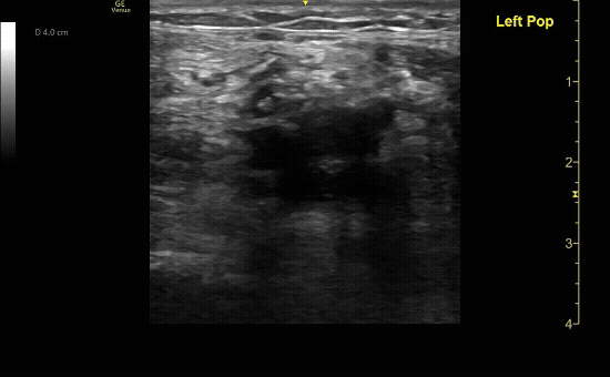
Ultrasound showing the popliteal trifurcation and compression just proximal to the popliteal vein. Be sure to show the vein before and after compression.
c/o Samson Frendo, MD
How to Scan:
POCUS 101: DVT Ultrasound Made Easy
Tips/Tricks/Pitfalls:
Always compress at an angle perpendicular to the vein to ensure compression is adequate
Be sure to increase your depth to confirm you are not imaging a superficial vein
If a patient has a large body habitus you can switch to the curvilinear probe for better visualization
You can increase sensitivity of the exam by performing systematic compressions of the entire lower extremity
For difficult exams, you can use your “non-probe” hand to compress the soft tissue from behind the leg (posteriorly🡪anteriorly) to assist in completely compressing the vessel you are investigating
Pathology:
Positive DVT ultrasound showing clot, and incomplete compression at the junction of the greater sapphenous vein and femoral vein.
c/o Samson Frendo, MD
Positive DVT ultrasound showing the bifurcation of the popliteal vein with incomplete compression of the popliteal vein.
c/o Matthew Hughes, MD
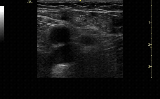
Proximal enlarged lymph node.
c/o Jordyn Cohen, MD
Key Literature:
Additional Resources:
Malin and Dawson iBook Volume 2, Chapter 12: DVT
ACEP Emergency Ultrasound Imaging Criteria Compendium Pages 50-55
AEUS Lecture by Frances Russell
Author: Niyi Soetan, MD (Class of 2026)
Reviewed by: David Murray, MD FDP-AEMUS