
Biliary
Learning Objectives:
Describe the indications and limitations of the bedside POCUS of the biliary tract.
Perform and review approved US views to evaluate the biliary tract.
Identify relevant ultrasound anatomy including the gallbladder, portal triad, inferior vena cava, and liver.
Recognize the relevant findings and pitfalls when evaluating for cholelithiasis and cholecystitis.
Indications:
Abdominal pain, right upper quadrant (RUQ) or epigastric
Suspicion for biliary colic, cholecystitis, cholangitis
Identification of symptomatic cholelithiasis
Identification of cholecystitis
Extended Indications:
Common bile duct pathology (dilatation and choledocholithiasis)
Liver pathology (masses, pneumobilia, hepatomegaly)
Portal vein abnormalities
Required Views:

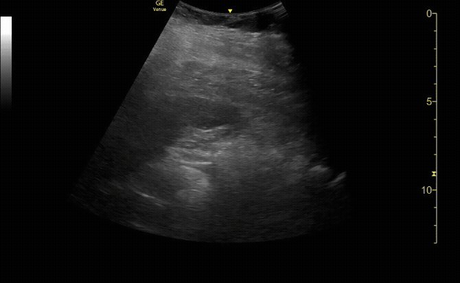
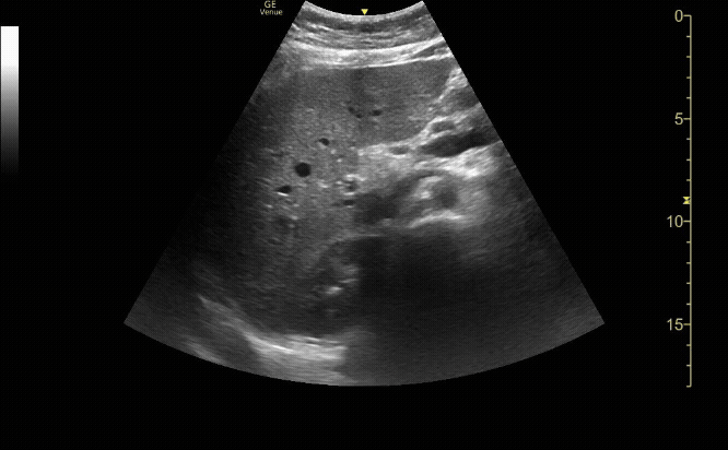
Gallbladder in long view
c/o Alex Alanis, MD
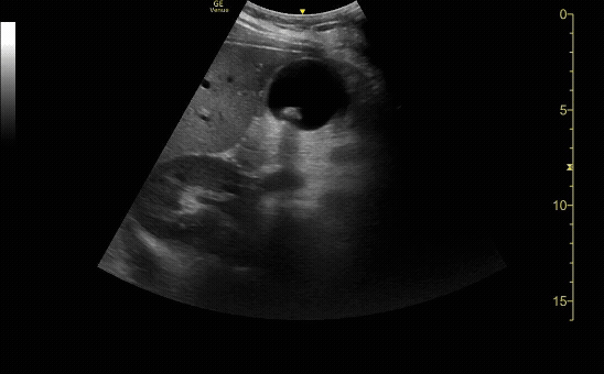
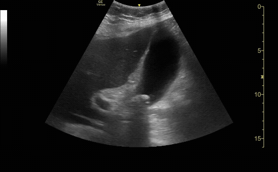
Gallbladder in short view
c/o Kristina Pfieffer, MD

Gallbladder wall measuring less than 0.3cm
c/o John Tucker, MD
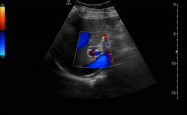
Common bile duct (if visualized) measuring <0.6cm with color flow seen in the portal vein.
c/o Jose Reyes, MD
How to Scan:
ACEP Sonoguide: Gallbladder Ultrasound
Core Ultrasound: Gallbladder US Basics
Core Ultrasound: Gallbladder US Pearls
POCUS 101: Abdominal Ultrasound Made Easy: Gallbladder
Tips/Tricks/Pitfalls:
Gallbladder visualization tips
When the gallbladder is in the long axis (the top of the exclamation point), the portal vein is in the short axis (the point of the exclamation point).
If there is difficulty visualizing the gallbladder in the supine position, roll the patient into the left lateral decubitus position.
Failure to identify the gallbladder may occur with chronic cholecystitis particularly when filled with stones, or, in the rare instances of gallbladder agenesis. Failure to identify the gallbladder should warrant additional diagnostic imaging such as radiologic ultrasound or computed tomography (CT).
The gallbladder may be confused with other fluid filled structures including the portal vein, the inferior vena cava, hepatic or renal cysts, or loculated collections of fluid. These can be more accurately identified with careful scanning in multiple planes and the use of color flow Doppler.
Gallstone tips
Gallstones are often mobile (move with patient positioning) and usually cause shadowing
Always visualize the neck of gallbladder for a single obstructing stone that may be cause of patient’s symptoms
Small gallstones may be overlooked or mistaken for gas in an adjacent loop of bowel. In questionable cases, gain settings should be optimized, the area should be scanned in several planes, and the patient should be repositioned to check for the mobility of gallstones.
Gallbladder polyps may be mistaken for gallstones. The former are non-mobile, do not shadow, and are adjacent and attached to the inner gallbladder wall. In certain circumstances polyps in the neck of the gallbladder can cause obstruction.
The presence of gallstones or other findings consistent with cholecystitis does not rule out the presence of other life-threatening causes of abdominal pain such as aortic aneurysm or myocardial infarction.
Gallbladder wall measurement tips
When measuring anterior gallbladder wall thickness - inflammation is not a uniform process and the wall should be measured at its thickest location. Measurement of gallbladder wall thickness should be made on the anterior wall, adjacent to the hepatic parenchyma in the transverse plane, to limit error secondary to oblique imaging and posterior acoustic enhancement artifact. Measurement of posterior gallbladder wall thickness may be inaccurate due to layered gallstones, acoustic enhancement from bile, and closely opposed loops of bowel.
Gallbladder wall thickening may not represent biliary pathology, but may be physiological, as in the contracted, post-prandial state, hypoproteinemia, liver disease, anasarca, and congestive heart failure.
Common bile duct tips
Doppler can be used to differentiate the CBD from the hepatic artery
Several studies have demonstrated increasing diameter with aging in patients without evidence of biliary disease. For this reason, many authorities consider that the normal common bile duct may increase by 1 mm for every decade of age.
Obstruction is the most common cause of CBD dilation: impacted stone, obstructing masses, or stricture.
The sensitivity for identifying common bile duct stones is low and often are only identified by the shadowing they cause or an abrupt narrowing of the common bile duct
Pathology:
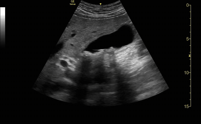
Gallbladder with stones and shadowing.
c/o Ryan Abbott, MD
Gallbladder in long view showing shadowing stones, thickened gallbladder wall, and pericholecystic fluid consistent with acute cholecystitis.
c/o Kevin Regis, MD
Gallbladder in short view showing WES (Wall, Echo, Shadow) sign indicating a gallbladder full of stones.
c/o Ginny Kim, MD
Gallbladder in long view demonstrating a stone-in-the-neck, can be considered equivalent to acute cholecystitis.
c/o Christopher Chang, MD
Key Literature:
Additional Resources:
Malin and Dawson iBook Volume 2, Chapter 15: RUQ
ACEP Emergency Ultrasound Imaging Criteria Compendium Pages 30-34
AEUS Lecture by Joseph Minardi
Bedside Ultrasound - Center of Ultrasound Education at Augusta University Pages 54-65
Core Ultrasound Gallbladder US with Dr. Fox
Emergency and Clinical Ultrasound: Basic Biliary Ultrasound wtih Dr. Lee LaRavia
Author: Priyanka Pradhan, MD (Class of 2026)
Reviewed by: Kyle Ackerman, MD