Aorta
Learning Objectives:
Describe indications, clinical algorithm, and limitations of POCUS in the evaluation of abdominal and thoracic aortic pathology
Perform clinical ultrasound protocols to evaluate the abdominal and thoracic aorta, including measurement techniques
Identify relevant US anatomy, including the aorta with major branches, the inferior vena cava, and the vertebral bodies
Recognize pathologic findings and pitfalls when evaluating for abdominal and thoracic aortic aneurysm and dissection
Integrate bedside aorta ultrasound for evaluation of acute abdominal/back pain management in the emergency department
Indications:
Evaluation of the abdominal aorta from the diaphragmatic hiatus to the aortic bifurcation for evidence of aneurysm
Extended Indications:
Abdominal aortic dissection
Thoracic aortic dissection
Presence of intraperitoneal free fluid when abdominal aortic aneurysm (AAA) is identified
Iliac, splenic, and other abdominal artery aneurysms
Presence of pericardial effusion, aortic regurgitation, and/or aortic root dilation (indirect signs) on bedside echocardiography when suspicion for thoracic aortic dissection is present
Required Views:
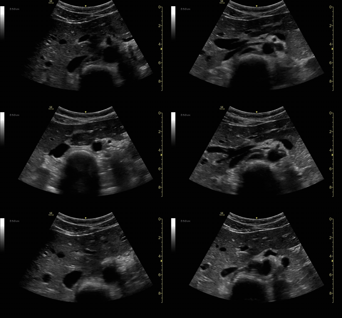
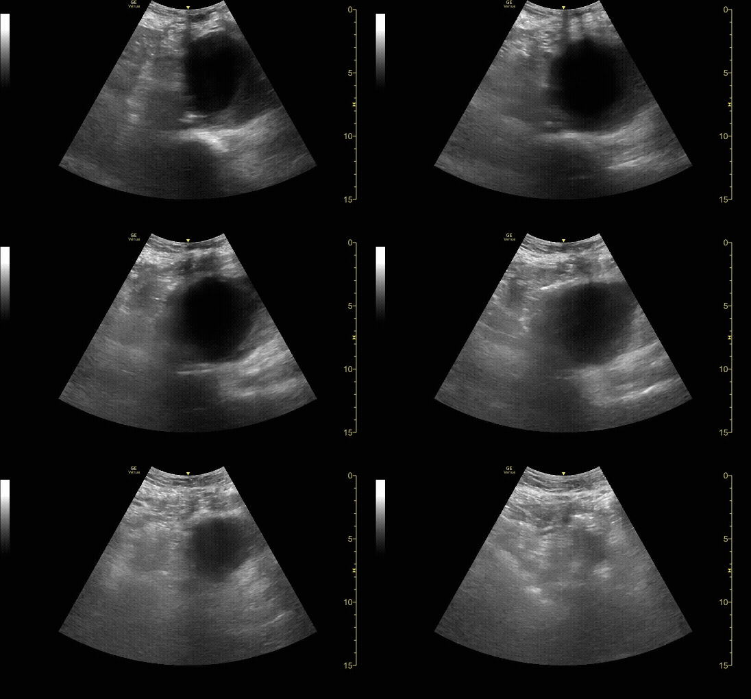
Proximal transverse (short) view of the aorta showing the celiac artery and SMA. Measure anterior-to-posterior proximally. Should measure <3cm.
c/o Taylor Wahrenbrock, MD
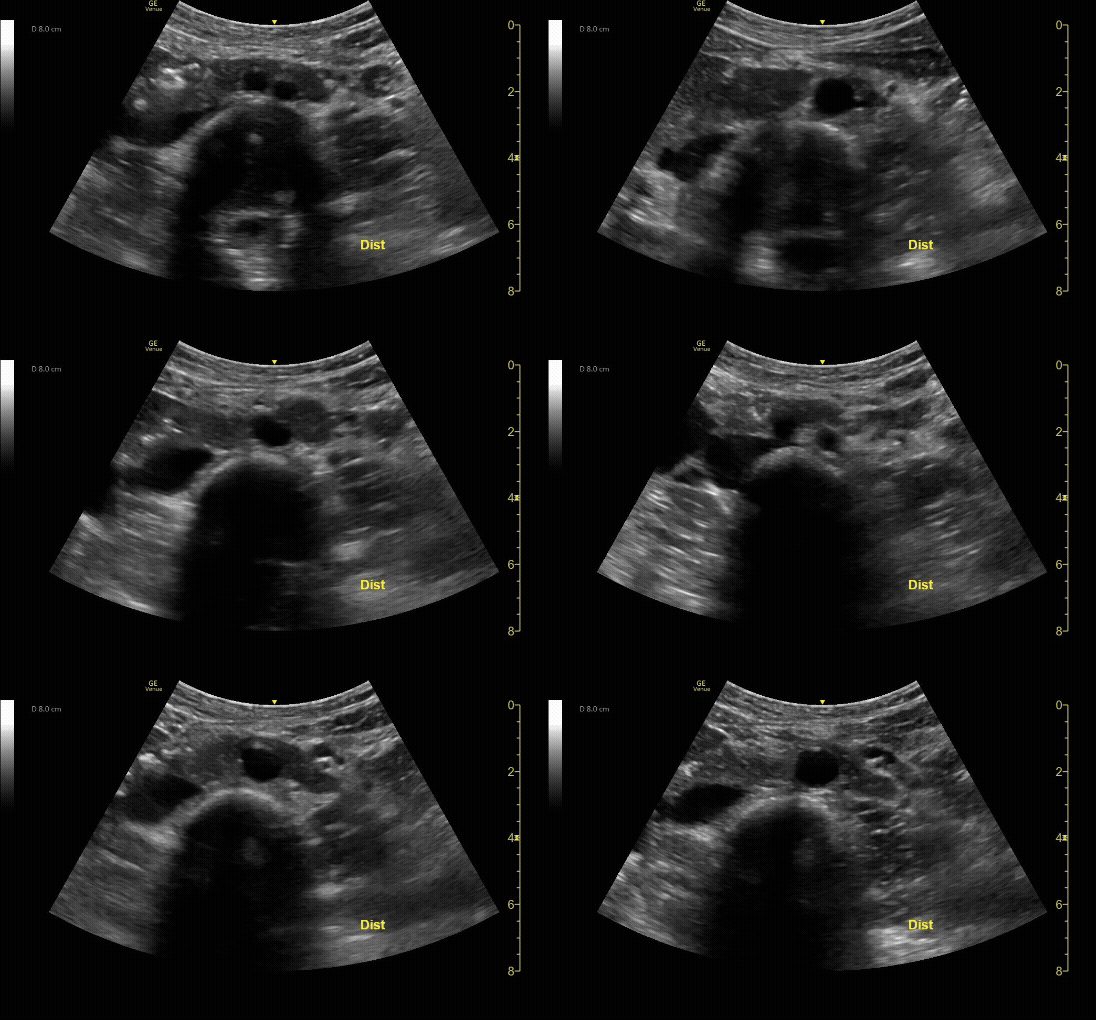
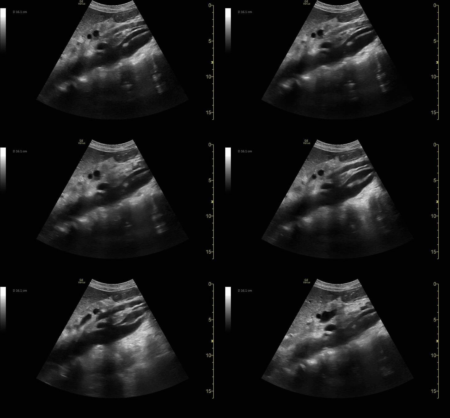
Distal transverse (short) view demonstrating the bifurcation. Measure anterior-to-posterior proximally. Should measure <3cm and taper distally.
c/o Taylor Wahrenbrock, MD
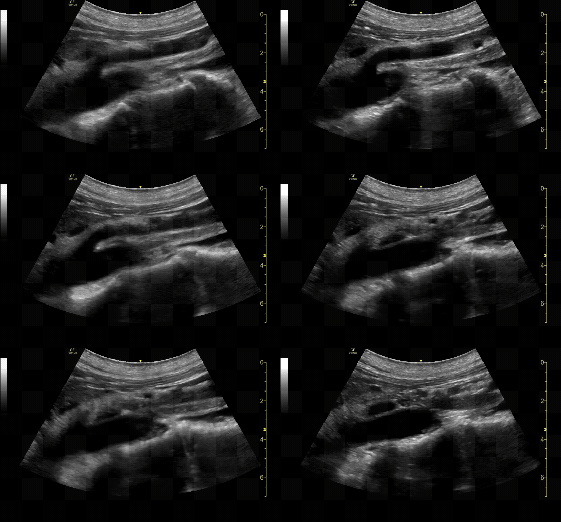
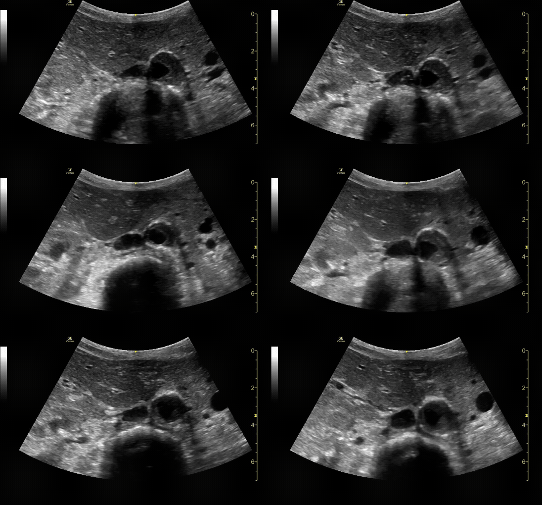
Proximal sagittal (long) view showing the SMA branching off of the aorta.
c/o Michael Dorritie, MD
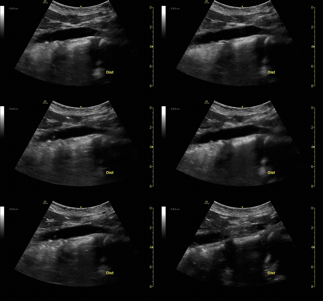
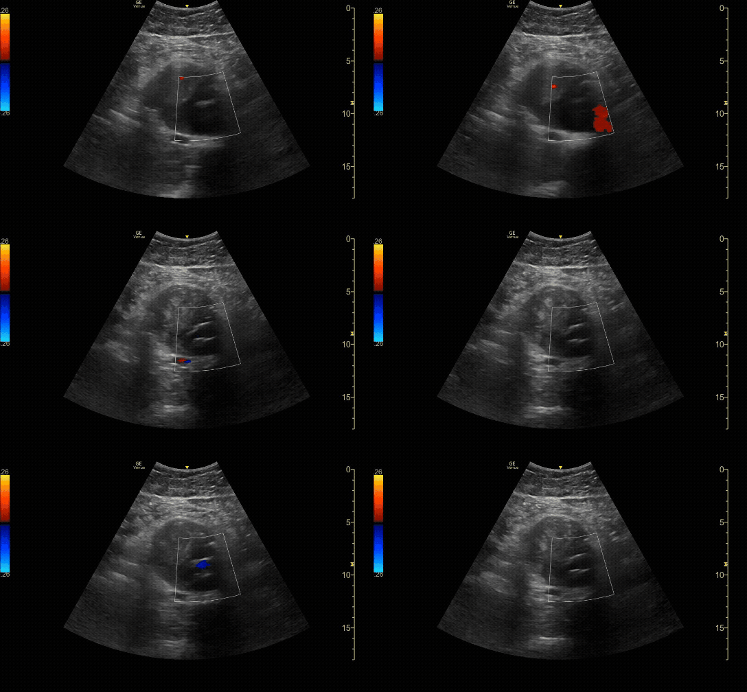
Distal sagittal (long) view at the bifurcation.
c/o Taylor Wahrenbrock, MD
How to Scan:
POCUS 101: Aorta Ultrasound Made Easy
Tips/Tricks/Pitfalls:
A small aortic diameter DOES NOT rule out rupture. Always combine the clinical picture with your US findings.
The aorta can be confused for the IVC. Make sure that you identify the bifurcation and branching vessels.
There can be clot in the aortic wall which can make the diameter appear falsely small. Be careful to make sure that you are measuring the true outer wall to outer wall.
Pathology:
Abdominal Aortic Aneurysm that measured 8cm in size.
c/o Ginny Kim, MD
Proximal sagittal (long) view showing a dissection flap.
c/o Sohaib Amjad, MD
Aorta in transverse (short) view with intramural thrombus.
c/o Alejandro Ruiz, MD
Transverse (short) view of an aorta after grafting with endoleak.
c/o Derek Hilgers, MD
Key Literature:
Additional Resources:
Malin and Dawson iBook Volume 1, Chapter 3: Aorta
ACEP Emergency Ultrasound Imaging Criteria Compendium Pages 2-5
Author: Niyi Soetan, MD (Class of 2026)
Reviewed by: Kyle Ackerman, MD

